

Comprehensive Cardiac
MRI Analysis
QIR-MR (for Quantified Imaging Resources) provides comprehensive quantitative cardiovascular MR analysis, from cardiac function and flow to tissue characterization and mapping.
Benefits
VALIDATED PERFORMANCE
≈90% CONTOUR OVERLAP
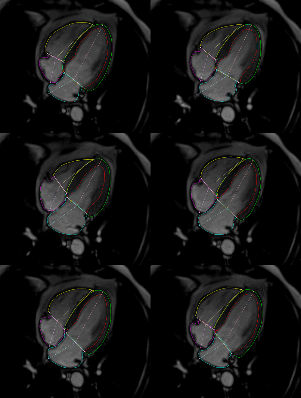
AI-based segmentation of the left ventricle, right ventricle and myocardium, validated against expert reference annotations.
AUTOMATED MYOCARDIAL SCAR DETECTION
Advanced DE-MRI / LGE analysis supports automatic detection, segmentation and quantification of myocardial scar and infarcted tissue.
MULTI-VENDOR VALIDATION
QIR-MR has been evaluated on cardiac MR datasets acquired across major MRI platforms and different field strengths, supporting robust performance in diverse clinical environments.
BACKED BY PEER-REVIEWED RESEARCH
QIR technologies are supported by extensive peer-reviewed research in cardiac segmentation, delayed-enhancement MRI and quantitative cardiovascular imaging.
Cardiac MR Workflow & Image Analysis
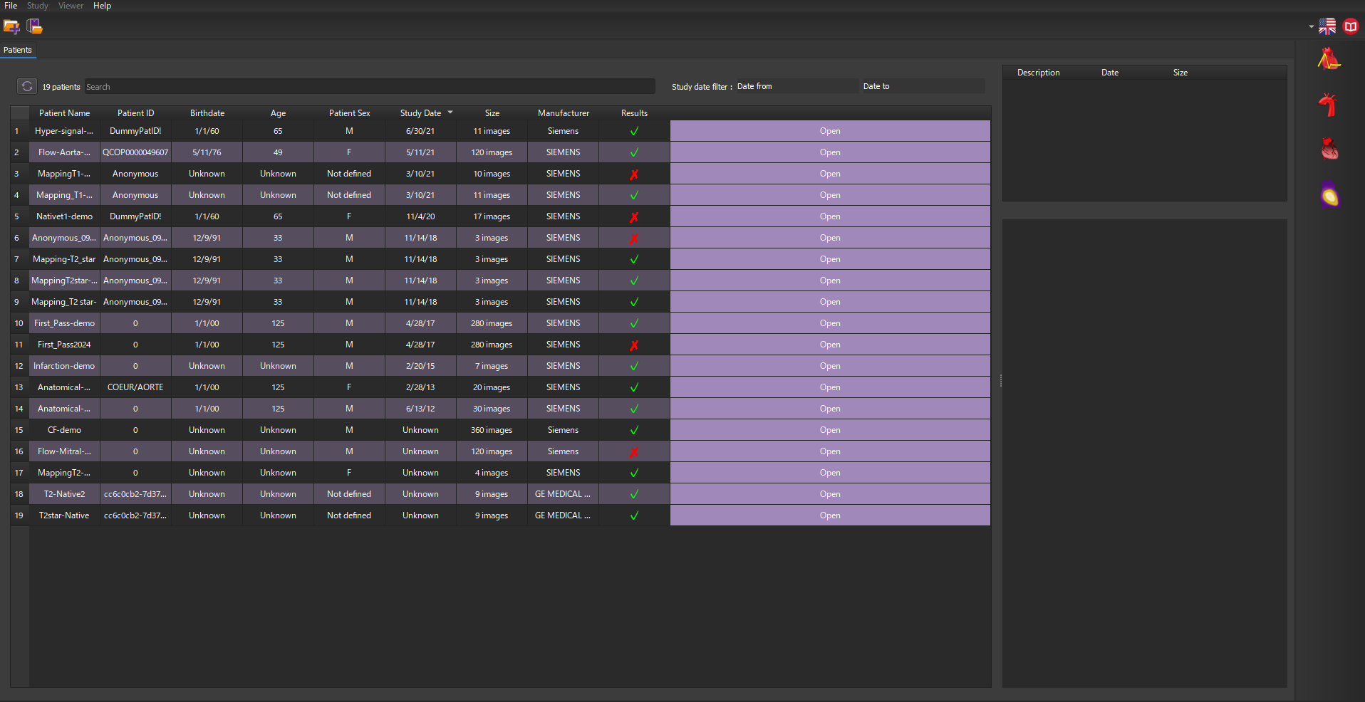
- Structured patient and examination management
- Automatic organization of cardiac MR series
- DICOM/PACS integration
- AI-assisted contour detection and propagation
- Advanced editing and measurement tools
- Multi-view visualization for efficient review
Cine Cardiac Module
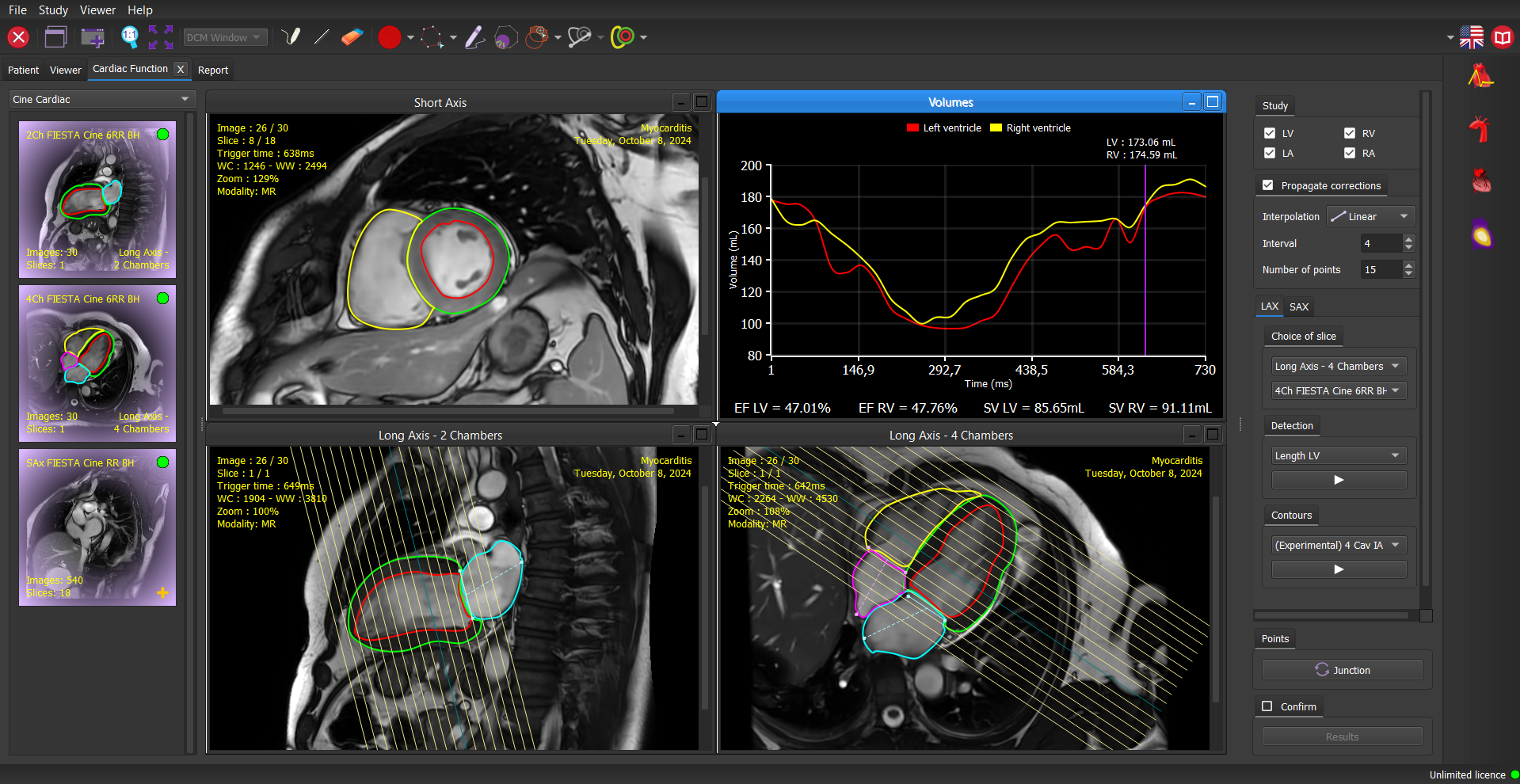
Automated ventricular function and myocardial strain analysis for cine cardiac MR.
- Cardiac function:
- Fast quantification of left and right ventricular function
- 4 Chambers analysisincluding atrial function assessment
- Papillary musclesand trabeculations detection
- Strain:
- New methods to study the 4 chambers strain
- Simplified strain
- Fast strain
- Diastolic and systolic contour propagation with improved detection time
- Automatic propagation of manual corrections
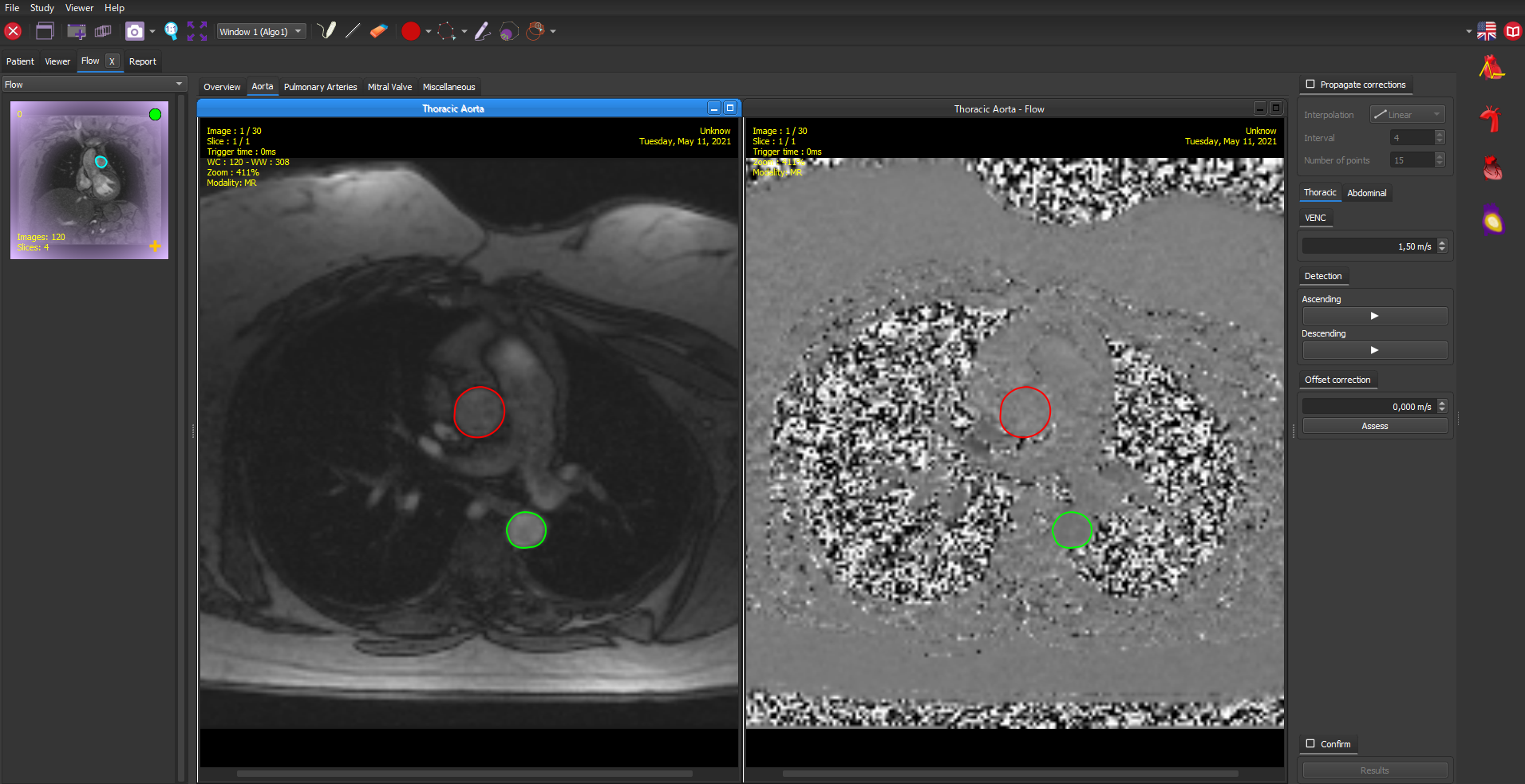
PC-MRI Module
Fast and robust algorithm providing automatic segmentation for the aorta
- 2D Flow:
- Proximal vessels & valve flow analysis
- Quantification of Cardiac output, Regurgitant fraction & Qp/Qs
- Compliance:
- Aorta length
Contrast Agent
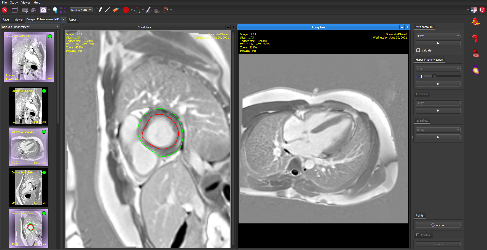
- DEMRI:
- 1st algorithm to detect false positives in myocardial infarction
- AI algorithm for myocardial contour detection
- nSD and FWHM methods for scar tissue quantification
- 17-segment Bull’s eye representation
- First Pass:
- Differentiate normally perfused from hypo-perfused areas (LV)
- Comparison between stress and rest images
- Motion correction algorithm
Mapping Module
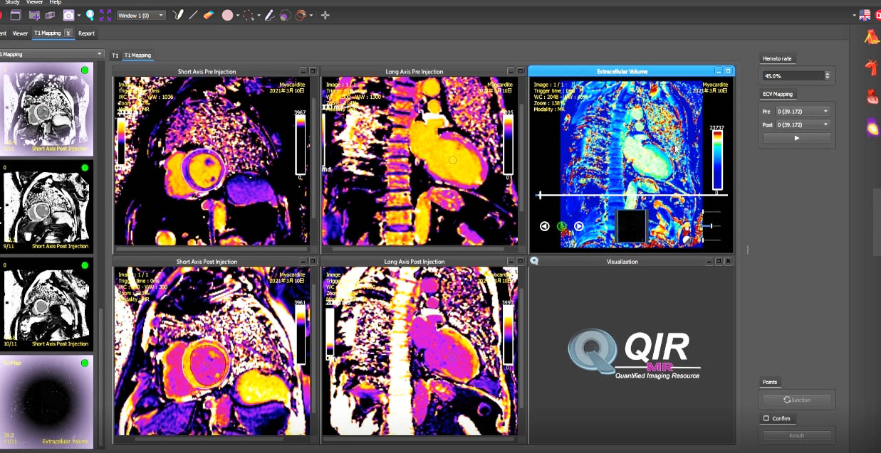
Global and regional analysis for SAX & LAX, color maps reconstruction from native images
- T1 Mapping:
- Calculate T1 relaxation times
- Extracellular volume fraction (ECV)
- T2 Mapping:
- Calculate T2 relaxation times
- T2* Mapping:
- Calculate T2* relaxation times
- Iron concentration in the myocardium and liver

7 impasse des Boussenots, 21800 Quetigny, France
+33 (0)3 80 79 11 14
contact@casis.fr
Support
Subscribe to Newsletter
- Join our subscribers list to get the latest news and special offers.
© 2026 - CASIS - CArdiac Simulation & Imaging Software | All Rights Reserved